Deepmala

Author name: aish.limelight

Blog

Can a Thyroid Nodule Be Treated Without Surgery?

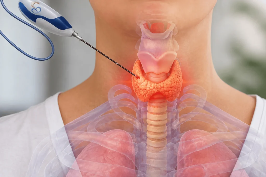
Can a Thyroid Nodule Be Treated Without Surgery? Yes — most benign thyroid nodules can now be treated without surgery For decades, the standard answer to a symptomatic thyroid nodule was surgical removal — either the nodule, part of the thyroid, or the entire gland. That is no longer the only option. Radiofrequency ablation (RFA) and microwave ablation (MWA) are non-surgical procedures that shrink thyroid nodules from the inside, preserving the thyroid gland entirely. Most patients with benign nodules are never told this option exists. This article explains what it is, who qualifies, what the results look like, and how it compares to surgery. What Is a Thyroid Nodule? A thyroid nodule is a discrete growth within the thyroid gland — a solid or fluid-filled lump that develops within the thyroid tissue. They are extremely common. Thyroid nodules are present in up to 65% of the general population when assessed with high-resolution ultrasound, though most are small and cause no symptoms. The majority of thyroid nodules are benign. Fewer than 5% are malignant. The clinical challenge is identifying which nodules require treatment and which can be safely monitored. How nodules are assessed — TIRADS and FNAC When a thyroid nodule is found on imaging, the standard assessment uses two tools: TIRADS (Thyroid Imaging Reporting and Data System) — TIRADS score determines whether FNAC is indicated. A standardised scoring system applied during ultrasound. It classifies nodules from TIRADS 1 (normal) to TIRADS 5 (high suspicion for malignancy) based on their ultrasound characteristics: composition, echogenicity, shape, margins, and echogenic foci. TIRADS score determines whether biopsy is indicated. FNAC (Fine Needle Aspiration Cytology) —  A thin needle is inserted under ultrasound guidance into the nodule. FNAC classifies the nodule as benign, indeterminate, suspicious, or malignant. This result determines the treatment pathway. Benign nodules confirmed on FNAC — particularly those causing symptoms due to their size — are the primary candidates for non-surgical ablation. When Does a Thyroid Nodule Need Treatment? Not every thyroid nodule requires intervention. Active surveillance — monitoring with periodic ultrasound — is appropriate for nodules that are small, stable, and asymptomatic. Treatment is indicated when any of the following apply: The nodule is causing compressive symptoms: difficulty swallowing, a sensation of pressure in the neck, hoarseness, or difficulty breathing The nodule is cosmetically significant — visible as a lump in the neck and causing distress The nodule is a functioning (autonomously hyperfunctioning) nodule producing excess thyroid hormone The nodule is growing on serial ultrasound — an increase of 20% or more in two dimensions FNAC results are indeterminate and the clinical picture warrants treatment over continued surveillance Historically, any of these presentations led to a surgical referral. For most patients, that is no longer necessary. What Is Thyroid RFA — Radiofrequency Ablation? Radiofrequency ablation (RFA) is a minimally invasive, non-surgical procedure that destroys thyroid nodule tissue using controlled heat delivered through a thin electrode needle. How the procedure works: Local anaesthetic is applied to the neck — no general anaesthesia is required A thin electrode needle is inserted into the nodule under continuous ultrasound guidance Radiofrequency energy is delivered through the needle tip, generating controlled heat within the nodule tissue The heat destroys the nodule cells while the surrounding normal thyroid tissue is preserved The procedure uses a ‘moving shot’ technique — the needle is repositioned within the nodule to ensure complete coverage Total procedure time is typically 30–60 minutes No incision. No stitches. No hospital stay required for most patients What happens after RFA: The treated nodule does not disappear immediately. Over the following weeks and months, the destroyed tissue is gradually absorbed and replaced by scar tissue. The nodule shrinks progressively: At 1 month: 30–40% volume reduction At 6 months: 50–80% volume reduction At 12 months: sustained reduction, with some nodules reducing by over 90% Compressive symptoms — difficulty swallowing, pressure — typically improve within weeks as the nodule begins to shrink. The thyroid gland remains intact and continues to function normally. What Is Microwave Ablation (MWA)? Microwave ablation uses microwave energy rather than radiofrequency to achieve the same result — controlled thermal destruction of nodule tissue. The mechanism differs slightly: MWA generates a larger ablation zone per application and reaches target temperature faster. Clinical outcomes are comparable between RFA and MWA. The choice between the two depends on nodule characteristics — size, location, vascularity — and operator expertise. Both are performed under ultrasound guidance, under local anaesthesia, without surgery. What Is Microwave Ablation (MWA)? Thyroid RFA / MWA Surgery (Thyroidectomy) Anaesthesia Local General Incision / scar None Yes — neck incision Hospital stay Day Care procedure 2–4 days Recovery time Recovery is immediate 1 week Thyroid preserved Yes Partial or complete removal Lifelong medication Usually not required Required if gland fully removed Nodule recurrence Extremely rare Minimal (gland removed) Risk to vocal nerves Very low Small but present Who Is a Candidate for Thyroid RFA? RFA is appropriate for most patients with benign, symptomatic thyroid nodules. Specifically, you are likely a good candidate if: Your nodule has been confirmed benign on FNAC The nodule is causing compressive symptoms or is cosmetically significant You want to preserve your thyroid gland and avoid lifelong thyroid hormone replacement You are not a suitable candidate for surgery due to other health conditions You had a previous thyroid surgery and have a recurrent nodule RFA is not appropriate for malignant nodules. Confirmed thyroid cancer requires surgical management in most cases, though RFA may have a role in specific scenarios — particularly recurrent or metastatic disease in inoperable patients, assessed on a case-by-case basis. Indeterminate FNAC results require individual clinical judgment. RFA is presented as an alternative to diagnostic surgery, but FNAC remains the recommended path. Key point on thyroid function after RFA: Because RFA targets only the nodule — not the surrounding gland — thyroid function is preserved in the vast majority of patients. Post-procedure thyroid hormone levels typically remain within normal range. This is one of the most significant advantages

Blog

Varicose Veins During and After Pregnancy: What’s Normal, What’s Not

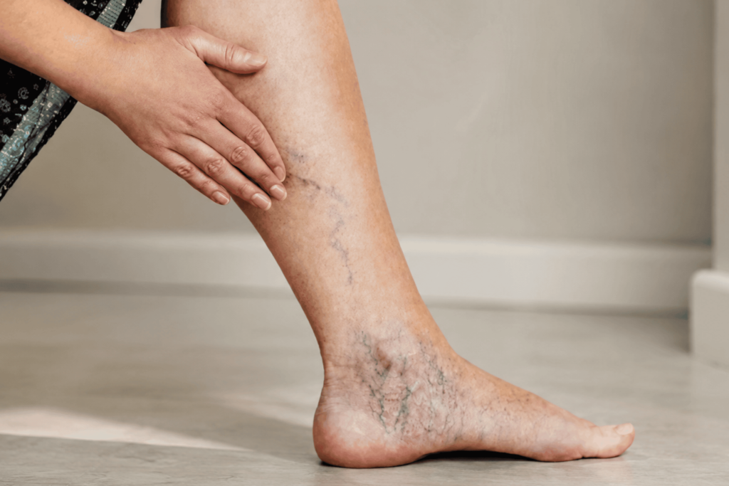
Varicose Veins During and After Pregnancy: What’s Normal, What’s Not Varicose veins are common in pregnancy. They are not always harmless. Most women are told to wait — that pregnancy-related varicose veins often resolve on their own after delivery. This is true for some. For others, the veins persist, worsen with subsequent pregnancies, and progress to a chronic venous insufficiency that becomes significantly harder to treat over time. This article explains what causes varicose veins in pregnancy, how to tell when they require attention, and what treatment looks like — during pregnancy, after delivery, and for women who were never told treatment was an option at all. Why Pregnancy Causes Varicose Veins Varicose veins develop when the valves inside the leg veins stop working properly. Healthy venous valves open to allow blood to flow upward toward the heart and close to prevent it from falling back down. When a valve fails, blood pools in the vein below it. The vein dilates under the pressure and becomes visible at the surface — this is a varicose vein. Pregnancy creates the ideal conditions for this to happen through three simultaneous mechanisms: 1. Increased blood volume Blood volume increases by 40–50% during pregnancy. The venous system — designed for a specific volume — is now carrying significantly more. The additional pressure strains the vein walls and the valves within them. 2. Hormonal changes Progesterone — which rises sharply during pregnancy — causes smooth muscle relaxation throughout the body, including the walls of blood vessels. Vein walls that would normally resist dilation become more compliant. Valves that were functioning adequately may begin to fail under the combined pressure of increased volume and reduced wall resistance. 3. Compression of pelvic veins The growing uterus compresses the inferior vena cava and the iliac veins — the major vessels that return blood from the legs to the heart. This compression raises the pressure in the leg veins further, accelerating valve failure. The compression is greatest in the third trimester and on the right side, which is why varicose veins often appear or worsen late in pregnancy. The result: venous pressure in the legs during pregnancy increases by approximately 3x compared to the non-pregnant baseline. For women with an underlying predisposition to venous insufficiency — genetic or otherwise — pregnancy is often the trigger that makes it visible. What Normal Looks Like — and What It Doesn’t Expected and generally self-resolving Mild varicose veins that appear during the second or third trimester and are limited to small, superficial veins. These often improve within 3–6 months postpartum as blood volume normalises, hormonal changes reverse, and pelvic compression is relieved. They may not disappear entirely, but significant improvement is common. Spider veins — the small, web-like red or purple vessels visible close to the skin surface — are extremely common in pregnancy and usually resolve without intervention. Not normal — requires assessment Any of the following warrant evaluation, not watchful waiting: Veins that are causing significant pain, heaviness, or aching in the legs — particularly pain that worsens through the day and improves with leg elevation Veins accompanied by swelling that is asymmetric — one leg notably more swollen than the other Skin changes around the ankle or lower leg: darkening (hyperpigmentation), thickening, or itching An area of the vein that becomes hard, red, and tender — this may indicate superficial thrombophlebitis, which requires urgent assessment Sudden, significant swelling in one calf accompanied by pain or warmth — this must be evaluated for deep vein thrombosis (DVT) immediately Vulval or perineal varicose veins, which develop in some pregnancies and require specialist assessment When to seek urgent assessment: One leg significantly more swollen than the other Calf pain with warmth and redness — possible DVT A section of varicose vein that is hard, red, and painful — superficial thrombophlebitis Any varicose veins that bleed — this can occur with minimal trauma and requires prompt management Varicose Veins After Pregnancy: What to Expect For most women, existing varicose veins improve in the first 3–6 months after delivery. The improvement is real but often incomplete — veins that were mildly visible before may become less prominent, but veins that were significantly dilated are unlikely to close entirely without treatment. Two patterns are clinically important: Veins that persist beyond 6 months postpartum If varicose veins are still present and symptomatic — causing aching, heaviness, swelling, or skin changes — at 6 months postpartum, spontaneous resolution is unlikely. These veins require treatment. The underlying valve failure does not reverse on its own. Veins that worsen with subsequent pregnancies Each pregnancy compounds the venous load. Women who develop mild varicose veins in their first pregnancy frequently find them more prominent after a second, and symptomatic after a third. The incremental valve damage accumulates. Treating varicose veins between pregnancies — when it is safe and technically straightforward — is more effective than treating advanced venous insufficiency after the family is complete. Treatment Options for Varicose Veins — Before and After Pregnancy During pregnancy Definitive treatment of varicose veins is not performed during pregnancy. The priorities are symptom management and complication prevention: Graduated compression stockings reduce venous pressure and improve symptoms significantly Leg elevation — particularly when resting — reduces pooling Regular walking maintains calf muscle pump function, which assists venous return Avoiding prolonged standing or sitting without movement These measures do not treat the underlying valve failure. They manage the load on the system while definitive treatment waits. After delivery — non-surgical treatment Once the postpartum period has elapsed and breastfeeding considerations are factored in, definitive treatment can be planned. The standard of care for varicose veins is now non-surgical. Endovenous Laser Treatment (EVLT) A thin laser fibre is inserted into the diseased vein through a small puncture — no surgical incision, no stripping. Under ultrasound guidance, the laser delivers controlled energy along the length of the vein, causing it to close. Blood is redirected to healthy veins. The treated vein is gradually absorbed by the

Blog

Why Do Fibroids Cause Back Pain?

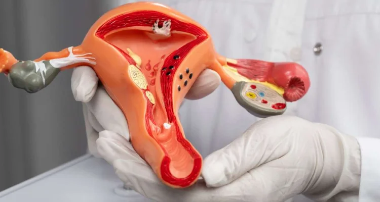
Can Fibroids Be Treated Without Surgery? Yes — fibroids can cause back pain. But the mechanism is specific, and most explanations online get it wrong. Fibroids cause back pain through direct mechanical pressure. As they grow, they can compress the nerves, muscles, and structures adjacent to the uterus — including the lumbar spine and the sciatic nerve. The result is pain that many women spend years attributing to posture, stress, or a ‘bad back.’ The fibroid is rarely the first suspect. It should be. What Is Happening Inside the Body The uterus sits in the pelvis, anterior to the rectum and posterior to the bladder. It is surrounded by a network of nerves, blood vessels, and connective tissue. When fibroids grow within or around the uterine wall, they increase the overall size and weight of the uterus significantly. A uterus without fibroids weighs approximately 60–80 grams. A fibroid-laden uterus can weigh 500 grams or more — the equivalent of a small melon occupying the pelvic cavity. This mass creates pressure in several directions: Posteriorly — toward the lumbar spine, sacrum, and sacral nerve roots Laterally — against the pelvic sidewall and uterosacral ligaments Inferiorly — downward pressure on the pelvic floor The back pain that results is not referred pain in the traditional sense. It is the direct consequence of physical compression. The Three Mechanisms That Cause Fibroid Back Pain 1. Nerve compression Posterior fibroids — those growing toward the back of the uterus — can compress the sacral nerve roots or the sciatic nerve directly. This produces a pain pattern that mimics sciatica: radiating pain down the buttock, the back of the thigh, and sometimes into the leg. Women with this pattern are frequently evaluated for disc herniation and spinal pathology before fibroids are considered. A pelvic ultrasound or MRI changes the picture immediately. 2. Uterosacral ligament tension The uterus is anchored to the sacrum by the uterosacral ligaments. As fibroids enlarge the uterus, they place sustained traction on these ligaments. The result is a deep, aching pain in the lower back and pelvis — typically worse during menstruation, when uterine contractions intensify the tension, and during prolonged standing or sitting. 3. Pelvic floor overload Fibroids increase the overall load on the pelvic floor musculature. The muscles compensate by contracting — producing tension that radiates into the lower back, hips, and glutes. This is often mistaken for muscular back pain and treated with physiotherapy, temporarily and incompletely. The source is not the muscle. The source is the mass creating the load. When the back pain is more likely fibroid-related: It is worse or began around the same time as heavy or prolonged periods It worsens during menstruation specifically It is accompanied by pelvic pressure or a sensation of fullness Standard physiotherapy or pain relief has not resolved it It is associated with frequent urination or difficulty emptying the bladder You have been diagnosed with fibroids, or haven’t been checked What Fibroid Back Pain Actually Feels Like Fibroid-related back pain is not always dramatic. It ranges from a persistent dull ache to sharp, debilitating pain depending on fibroid size, location, and whether nerve compression is involved. Common descriptions include: A constant low-grade ache across the lower back, not associated with a specific movement or injury A heaviness or dragging sensation in the pelvis that radiates into the back Pain that intensifies during menstruation and eases somewhat afterward — then returns Sharp, shooting pain down one or both legs (if sciatic nerve compression is present) Pain that worsens after prolonged sitting or standing, and is not relieved by rest The cyclical pattern — pain that correlates with the menstrual cycle — is one of the clearest indicators that the pelvis, not the spine, is the source. Why Is This So Often Missed? Back pain is one of the most common complaints in clinical medicine. The default investigations — X-ray, MRI of the lumbar spine, orthopaedic referral — look at the spine and miss the pelvis entirely. Fibroids take an average of four years to diagnose from the onset of symptoms. Part of that delay is the fragmentation of care: a woman with back pain sees an orthopaedic surgeon or physiotherapist. A woman with heavy periods sees a gynaecologist. The two symptom clusters are rarely connected unless someone thinks to look at both. A pelvic ultrasound — a simple, inexpensive investigation — would identify most clinically significant fibroids. It is not routinely included in the workup for back pain. It should be, in women with any accompanying pelvic symptoms. Other Fibroid Symptoms That Accompany Back Pain Back pain from fibroids rarely presents in isolation. If you have lower back pain and any of the following, fibroids should be investigated: Heavy menstrual bleeding — soaking through pads or tampons, passing clots Periods lasting longer than 7 days Pelvic pain or cramping outside of menstruation Bloating or a distended lower abdomen Frequent urination or difficulty fully emptying the bladder Pain during intercourse Difficulty conceiving The presence of multiple symptoms in this cluster makes fibroid-related back pain significantly more likely. How Fibroids Are Diagnosed If fibroids are suspected based on symptoms, the initial investigation is a pelvic ultrasound. This is performed transabdominally or transvaginally and will identify the presence, size, number, and general location of fibroids. When more detail is needed — particularly for treatment planning — an MRI of the pelvis is the gold standard. MRI defines fibroid location precisely (submucosal, intramural, subserosal, pedunculated), assesses vascularity, and rules out other pelvic pathology that could be contributing to symptoms. Neither investigation is invasive. Both are widely available. What Treating the Fibroids Does to the Back Pain Back pain caused by fibroid compression resolves when the fibroids are treated. The mechanism is straightforward — remove the pressure, remove the pain. Treatment options include: Uterine Fibroid Embolization (UFE) A non-surgical procedure in which the blood supply to the fibroids is blocked through a catheter inserted at the wrist or groin. Without blood supply, fibroids shrink by

Blog

Can Fibroids Be Treated Without Surgery?

Can Fibroids Be Treated Without Surgery? If you have been told that surgery is your only option for fibroids — you have not been given the full picture. A non-surgical treatment for uterine fibroids has existed for over two decades. It is called Uterine Fibroid Embolization, or UFE. Most women with fibroids have never heard of it. This article explains what it is, who it is right for, and how it compares to the surgical options you may already have been offered. What Are Uterine Fibroids? Uterine fibroids are non-cancerous growths that develop within the muscle wall of the uterus. They are extremely common — fibroids affect approximately 1 in 3 women at some point in their lives, and up to 70% of women will develop at least one fibroid by the age of 50. Fibroids range in size from a few millimetres to several centimetres. Some women have a single fibroid. Others have multiple. Their location within the uterus — submucosal, intramural, or subserosal — determines what symptoms they cause. Common symptoms include: Heavy or prolonged menstrual bleeding Pelvic pain or pressure Frequent urination Lower back pain Pain during intercourse Bloating or a visibly enlarged abdomen Not all fibroids cause symptoms. When they do, the severity varies widely. The decision to treat — and how — depends on the symptoms, the fibroid’s size and location, and the patient’s fertility plans. The Standard Treatment Options for Fibroids When a woman is diagnosed with symptomatic fibroids, she is typically presented with three options: 1. Hysterectomy Surgical removal of the uterus. This is the most commonly recommended treatment for fibroids in India. It is definitive — fibroids cannot recur because the uterus is removed. However, it is also major surgery. It requires general anaesthesia, a hospital stay of 2–5 days, and a recovery period of 4–8 weeks. And it permanently ends the possibility of pregnancy. 2. Myomectomy Surgical removal of individual fibroids while preserving the uterus. This is the standard recommendation for women who wish to maintain fertility. It can be done as open surgery, laparoscopically, or hysteroscopically, depending on fibroid location. Recovery is shorter than hysterectomy but still involves surgery, anaesthesia, and downtime. Fibroids can also recur — studies show recurrence rates of 15–30% within 3 years. 3. Medication Hormonal therapies such as GnRH analogues can temporarily reduce fibroid size and control bleeding. They do not eliminate fibroids. Once the medication stops, fibroids typically return to their previous size. Medication is usually used short-term before surgery, not as a standalone long-term solution. What most women are not told about is the fourth option. What Is UFE — Uterine Fibroid Embolization? Uterine Fibroid Embolization (UFE) is a minimally invasive, non-surgical procedure that treats fibroids by cutting off their blood supply. Fibroids are highly dependent on blood flow to survive and grow. UFE works by blocking the blood vessels that feed the fibroids. Without that supply, the fibroids shrink — and symptoms resolve. How the procedure works: A thin catheter is inserted through a small puncture in the wrist or groin — no incision, no stitches Using live X-ray imaging (fluoroscopy), the catheter is guided to the uterine arteries Tiny particles are injected to block the blood supply to the fibroids The procedure takes approximately 60–90 minutes Most patients stay one night in hospital and return to normal activity within 7–10 days What happens to the fibroids after UFE: The fibroids do not disappear overnight. Over the following 3–6 months, they shrink progressively as they lose their blood supply. Clinical data shows: Fibroid volume reduction of 50–60% at 6 months Significant improvement in heavy bleeding in over 85% of patients Sustained symptom relief in the majority of patients at 5-year follow-up The uterus is preserved. There are no surgical incisions. No general anaesthesia is required. UFE vs Hysterectomy vs Myomectomy: A Clinical Comparison UFE Hysterectomy Myomectomy Surgery required No Yes (major) Yes General anaesthesia No Yes Yes Hospital stay 1 night 3–5 days 2–4 days Recovery time 7–10 days 4–8 weeks 2–6 weeks Uterus preserved Yes No Yes Fibroid recurrence Low None (uterus removed) 15–30% at 3 years Fertility preserved Possible No Yes Who Is UFE Right For? UFE is appropriate for most women with symptomatic fibroids who want to avoid surgery. Specifically, UFE is a strong option if you: Have heavy periods, pelvic pain, or pressure that is affecting your quality of life Have been told you need a hysterectomy and want an alternative Want to preserve your uterus Cannot tolerate major surgery due to other health conditions Have multiple fibroids that make myomectomy complex UFE may not be suitable in certain situations. Your interventional radiologist will assess candidacy based on: Fibroid location and vascularity (assessed on MRI) Whether you are planning a pregnancy in the near future Presence of suspected malignancy — UFE is not appropriate for cancerous lesions Specific fibroid subtypes such as pedunculated submucosal fibroids A pre-procedure MRI is standard. It maps the fibroids precisely and rules out conditions that would change the treatment plan. Can You Get Pregnant After UFE? This is the question most women ask first — and the answer requires nuance. UFE is not the first-line recommendation for women who are actively planning a pregnancy in the immediate term. Myomectomy remains the standard recommendation in that setting, because it allows direct fibroid removal while preserving fertility pathways. That said, pregnancies after UFE are documented. Published case series show successful pregnancies and live births following UFE. The decision depends on fibroid burden, age, and the specific fertility picture — and should be made in consultation with both an interventional radiologist and a gynaecologist. For women who have completed their family or are not planning pregnancy, UFE offers uterine preservation without this consideration. Frequently Asked Questions Is UFE painful? There is post-procedural cramping and discomfort in the first 24–48 hours as the fibroids respond to reduced blood supply. This is managed with medication in hospital. Most patients describe it as significantly more manageable than anticipated, and

Scroll to Top